Posts

Showing posts with the label RadiologyCases

Preiser Disease (Idiopathic Avascular Necrosis of the Scaphoid): A Comprehensive Clinical, Radiological, and Therapeutic Review for Global Medical Practice

Image
  Keywords: Preiser disease, scaphoid avascular necrosis, idiopathic scaphoid AVN, wrist pain diagnosis, scaphoid collapse, proximal pole necrosis, radiology imaging of wrist, orthopedic hand surgery, MRI scaphoid necrosis, differential diagnosis of wrist pain Introduction Preiser disease is a rare but clinically significant form of idiopathic avascular necrosis (AVN) of the scaphoid , first described by Georg Preiser in 1910. Unlike post-traumatic scaphoid osteonecrosis, Preiser disease occurs in the absence of preceding fracture or trauma , posing diagnostic challenges for clinicians and radiologists alike. The scaphoid bone plays a central biomechanical role in wrist stability , bridging the proximal and distal carpal rows. Compromise of its vascular supply—especially in the proximal pole—can lead to progressive collapse, degenerative arthritis, chronic pain, and significant functional disability . Clinical Case Patient Profile: Age: 21 years Sex: Female Chief ...

Auricular Cartilage Calcification and Adrenal Insufficiency: Pathophysiology, Imaging Features, Diagnosis, and Clinical Implications – A Comprehensive Expert Review

Image
  Keywords: Auricular cartilage calcification, adrenal insufficiency, autoimmune hypophysitis, CT imaging, skull radiography, hypotensive shock, endocrine emergency, radiologic diagnosis, pituitary disorders, adrenal crisis Introduction Auricular cartilage calcification is an exceptionally rare radiologic and clinical entity, often underrecognized and misdiagnosed. When it occurs in conjunction with adrenal insufficiency, it represents a potentially life-threatening endocrine emergency requiring immediate diagnosis and treatment. This article presents a detailed case-based discussion of auricular cartilage calcification associated with autoimmune hypophysitis-induced adrenal insufficiency , integrating pathophysiology, epidemiology, clinical presentation, imaging findings, differential diagnosis, diagnostic strategies, treatment, and prognosis , supported by the most authoritative and up-to-date medical literature. The importance of recognizing auricular cartilage calcificati...

Osmotic Demyelination Syndrome (Central Pontine Myelinolysis): Pathophysiology, Imaging Features, Diagnosis, and Clinical Management

Image
  Keywords: Osmotic Demyelination Syndrome, Central Pontine Myelinolysis, Hyponatremia Correction, MRI Findings, Diffusion-Weighted Imaging, Extrapontine Myelinolysis, Neurologic Complications Introduction Osmotic Demyelination Syndrome (ODS), classically known as Central Pontine Myelinolysis (CPM) , is a rare but devastating neurologic disorder characterized by non-inflammatory demyelination , most commonly affecting the central pons. First described in malnourished and alcoholic patients, ODS is now recognized as a complication of rapid correction of chronic hyponatremia and other abrupt osmotic shifts. Despite advances in neuroimaging and critical care, ODS remains associated with significant morbidity and mortality. Early recognition, appropriate sodium correction strategies, and timely imaging—particularly diffusion-weighted MRI —are essential to improving outcomes. This article provides a comprehensive, literature-based review of ODS, integrating pathophysiology, epi...

Intestinal Malrotation: Pathophysiology, Imaging Features, Diagnosis, and Management

Image
  Introduction Intestinal malrotation is a rare but clinically significant congenital anomaly of intestinal rotation and fixation. Although typically diagnosed in infancy, it can occasionally present in adulthood with atypical features such as chronic abdominal pain, intermittent vomiting, or even life-threatening midgut volvulus. This article provides a comprehensive overview based on the latest global literature , while integrating illustrative radiographic and CT images for accurate interpretation. Case Summary A 59-year-old woman presented with chronic abdominal pain . Radiographs and CT scans revealed abnormal positioning of the small and large intestines, with features consistent with intestinal malrotation . Imaging Findings from the Case [Figure 1] Simple abdomen X-ray (A: Erect, B: Supine): Dilated colonic loops clustered in the left hemiabdomen , with small bowel loops predominantly in the right hemiabdomen . Interpretation: Suggestive of abnormal intestinal pos...

The Hot Quadrate Sign in Superior Vena Cava Obstruction: A Radiological and Clinical Guide

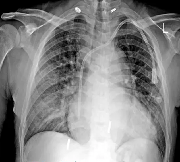
Image
  The Hot Quadrate Sign in Superior Vena Cava Obstruction: A Radiological and Clinical Guide El signo del cuadrado caliente en la obstrucción de la vena cava superior: una guía radiológica y clínica 상대정맥 폐쇄 시 나타나는 뜨거운 사각 징후: 방사선 및 임상 가이드 Keywords : Superior vena cava syndrome, hot quadrate sign, hepatic segment IV enhancement, thoracic CT, venous obstruction, dialysis catheter complication, radiological diagnosis, interventional radiology Introduction A 47-year-old man with a history of end-stage renal disease on dialysis presented with the sudden onset of dyspnea. A chest radiograph and subsequent CT angiography revealed a rare but diagnostically significant finding: superior vena cava (SVC) obstruction accompanied by the hot quadrate sign on liver imaging. This case presents a classic example of venous obstruction leading to collateral circulation and retrograde portal perfusion. Clinical Case Overview Patient : 47-year-old male Chief complaint : Acute shortness of ...