The Hot Quadrate Sign in Superior Vena Cava Obstruction: A Radiological and Clinical Guide
The Hot Quadrate Sign in Superior Vena Cava Obstruction: A Radiological and Clinical Guide
Keywords: Superior vena cava syndrome, hot quadrate sign, hepatic segment IV enhancement, thoracic CT, venous obstruction, dialysis catheter complication, radiological diagnosis, interventional radiology
Introduction
A 47-year-old man with a history of end-stage renal disease on dialysis presented with the sudden onset of dyspnea. A chest radiograph and subsequent CT angiography revealed a rare but diagnostically significant finding: superior vena cava (SVC) obstruction accompanied by the hot quadrate sign on liver imaging. This case presents a classic example of venous obstruction leading to collateral circulation and retrograde portal perfusion.
Clinical Case Overview
-
Patient: 47-year-old male
-
Chief complaint: Acute shortness of breath
-
Medical history: End-stage renal disease on hemodialysis
Imaging Findings:
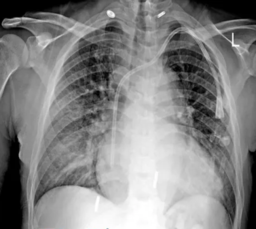 |
| Figure 1: Chest X-ray reveals an enlarged cardiac silhouette and diffuse interstitial infiltrates indicative of pulmonary edema. |
 |
| Figure 2: Thoracic CT angiography shows complete occlusion of the SVC with collateral thoracic venous drainage and intense enhancement of hepatic segment IV, forming the “hot quadrate sign.” |
The Hot Quadrate Sign: Radiologic Pearl
The hot quadrate sign refers to the intense contrast enhancement of segment IV (the quadrate lobe) of the liver during contrast-enhanced CT, typically associated with SVC obstruction.
Mechanism:
When the SVC is obstructed, normal venous return from the upper body is rerouted through alternative pathways. One of these collaterals includes veins of the anterior abdominal wall that eventually drain into the left portal vein, particularly enhancing segment IV of the liver. This focal hepatic enhancement gives rise to the “hot quadrate sign.”
-
Key implication: It is a pathognomonic sign indicating chronic or acute SVC obstruction and may be seen even in asymptomatic cases with central venous thrombosis.
Etiology of SVC Obstruction
SVC obstruction can be classified based on its origin as malignant, iatrogenic, or inflammatory.
Common Causes:
-
Iatrogenic (Most common in dialysis patients):
-
Central venous catheters
-
Hemodialysis lines
-
Pacemaker wires
-
-
Malignancies:
-
Lung cancer (especially small cell)
-
Non-Hodgkin lymphoma
-
Metastatic mediastinal tumors
-
-
Other:
-
Infections (e.g., tuberculosis)
-
Fibrosing mediastinitis
-
Pathophysiology
SVC obstruction increases venous pressure in the upper body, leading to:
-
Facial and neck swelling
-
Engorged neck and chest veins
-
Headache, dizziness, and visual disturbances
-
Dyspnea and orthopnea
-
In chronic settings, the development of extensive collateral venous pathways, including the azygos system and abdominal wall veins
The hot quadrature sign arises due to:
-
Retrograde flow through the paraumbilical veins and the left portal vein
-
Localized contrast pooling in segment IV of the liver
Clinical Presentation
Patients with SVC syndrome may present with:
| Symptom | Explanation |
|---|---|
| Dyspnea | Due to impaired venous return and pulmonary congestion |
| Facial Swelling | Venous engorgement of the upper body |
| Jugular Distension | Increased central venous pressure |
| Cyanosis | Impaired oxygenation |
| Hepatic Enhancement | Via an alternative venous inflow into the portal system |
In this case, the acute dyspnea and radiographic signs of pulmonary edema were compounded by a thrombosed dialysis catheter, highlighting the importance of catheter-related complications in SVC syndrome.
Imaging Features
Chest X-ray:
-
Enlarged cardiac silhouette (Cardiomegaly)
-
Diffuse interstitial opacities (Suggestive of pulmonary edema)
-
The tip of the dialysis catheter is positioned in the right atrium
CT Angiography:
-
Occluded SVC with central thrombus
-
Extensive collateral thoracic and abdominal venous enhancement
-
Prominent enhancement of hepatic segment IV (Hot Quadrate Sign)
Differential Diagnosis
| Diagnosis | Imaging Clues | Key Differentiators |
|---|---|---|
| Pulmonary embolism | Filling defects in pulmonary arteries | No hepatic enhancement |
| Pancoast tumor | Apical lung mass, rib destruction | No liver findings |
| Mixing artifact | Transient liver enhancement near IVC | Segment IV is not selectively enhanced |
| Hepatocellular carcinoma | Mass with arterial enhancement | Persistent enhancement beyond the arterial phase |
| SVC Obstruction with Hot Quadrate Sign | SVC occlusion, hepatic segment IV enhancement | Pathognomonic pattern |
Treatment Options
The management of SVC obstruction depends on the underlying cause and severity of symptoms.
1. Medical Management:
-
Anticoagulation: For thrombotic causes
-
Diuretics: To manage pulmonary edema
-
Steroids: If due to malignancy or inflammation
2. Interventional Radiology:
-
Thrombolysis
-
Balloon angioplasty
-
Stent placement
3. Surgical Management:
-
Tumor resection
-
Bypass grafting for refractory cases
In catheter-induced obstruction, removal or repositioning of the catheter is essential.
Prognosis
-
Favorable when the cause is iatrogenic and treated early
-
Poorer prognosis in cases of malignant obstruction or delayed diagnosis
-
Hot quadrate sign may persist for some time even after resolution of SVC obstruction, due to residual venous rerouting
Quiz
1. On the chest radiograph, the heart appears to be enlarged.
A. True
B. False
2. Which of the following best explains the diffuse interstitial opacities seen on the chest radiograph?
A. Pulmonary embolism
B. Lymphangitic carcinomatosis
C. Interstitial lung disease
D. Pulmonary edema
3. Is there evidence of a central pulmonary embolus on the CT scan?
A. Yes
B. No
4. In the general population, what is the most common cause of superior vena cava obstruction as seen in this case?
A. Lung cancer
B. Lymphoma
C. Iatrogenic causes (e.g., catheters)
D. Metastatic disease
5. Which liver segment is classically involved in the hot quadrate sign on CT imaging?
A. Segment II
B. Segment III
C. Segment IV
D. Segment V
6. What is the final diagnosis in this patient?
A. Pulmonary embolism
B. Pancoast tumor
C. Mixing artifact
D. Superior vena cava obstruction with hot quadrate sign
Answer & Explanation
1. Correct Answer: A. True, Explanation: The chest X-ray in this case reveals a widened mediastinal silhouette with an enlarged cardiac shadow, consistent with cardiomegaly, which may be due to volume overload in a patient with end-stage renal disease.
2. Correct Answer: D. Pulmonary edema, Explanation: The bilateral diffuse interstitial and alveolar opacities on the radiograph are indicative of pulmonary edema, which is common in patients with volume overload due to renal failure and dialysis-dependent states.
3. Correct Answer: B. No, Explanation: Although dyspnea is a shared symptom, no thrombus or filling defect is seen in the main or lobar pulmonary arteries. Instead, findings show SVC occlusion, making pulmonary embolism unlikely in this case.
4. Correct Answer: C. Iatrogenic causes (e.g., catheters), Explanation: In recent decades, particularly in dialysis patients, central venous catheters have become the leading cause of SVC obstruction. In this case, the tip of the dialysis catheter is thrombosed at the level of the right atrium/SVC junction.
5. Correct Answer: C. Segment IV, Explanation: The quadrate lobe (segment IV) of the liver receives redirected venous blood through the paraumbilical and left portal veins during SVC obstruction, resulting in localized enhancement known as the hot quadrate sign.
6. Correct Answer: D. Superior vena cava obstruction with hot quadrate sign., Explanation: CT angiography shows thrombosed SVC, extensive collateral venous circulation, and a hyperenhanced segment IV of the liver, consistent with the classic imaging findings of SVC obstruction with hot quadrate sign. No evidence of tumor or embolism is found.
Conclusion
The hot quadrate sign is a striking radiologic indicator of SVC obstruction, providing critical diagnostic insight in cases of unexplained dyspnea or suspected central venous thrombosis. In dialysis patients, vigilance for catheter-related complications is vital. Early identification via CT imaging allows for timely intervention, potentially averting serious outcomes.
References (IEEE Style)
-
J. Y. Lee et al., “CT findings of SVC syndrome: correlation with the extent and cause of obstruction,” AJR Am J Roentgenol, vol. 193, no. 2, pp. 757–764, 2009.
-
C. B. Smith et al., “Superior vena cava syndrome: diagnosis and treatment,” Cancer J Clin, vol. 58, pp. 38–52, 2008.
-
T. Matsumoto et al., “Hot quadrate lobe sign: A clue to SVC obstruction,” Radiology, vol. 262, no. 3, pp. 947–955, 2012.
-
M. D. Rosado-de-Christenson and J. W. Abbott, “From the radiologic pathology archives: SVC syndrome,” Radiographics, vol. 27, pp. 937–960, 2007.
-
R. A. Muller et al., “CT imaging of thoracic venous obstruction,” J Comput Assist Tomogr, vol. 30, pp. 564–572, 2006.
-
A. Y. Kim et al., “Collateral pathways in thoracic venous obstruction,” Korean J Radiol, vol. 10, no. 2, pp. 137–144, 2009.
-
S. J. Im et al., “Characteristic hepatic enhancement: hot quadrate lobe sign in CT,” Clin Imaging, vol. 36, pp. 478–480, 2012.


Comments
Post a Comment