Leukodystrophy on Brain CT and MRI: A Rare but Devastating White Matter Disease
Leukodystrophy on Brain CT and MRI: A Rare but
Devastating White Matter Disease
Introduction
Leukodystrophies are a group of rare,
progressive genetic disorders that affect the white matter of the brain and
spinal cord. Characterized by abnormal development or destruction of myelin, the
protective sheath surrounding nerve fibers, these diseases result in a spectrum
of neurological impairments.
This article focuses on the brain CT and
MRI findings of leukodystrophy, illustrated by a 30-year-old male patient. The
radiological features are discussed along with pathophysiological mechanisms,
epidemiology, clinical manifestations, and the latest therapeutic strategies.
Case Presentation

A
30-year-old man underwent brain CT for evaluation of progressive cognitive
decline. Compared to normal axial slices through the midbrain, the patient's
images revealed:
·
Global ventricular enlargement and CSF space expansion (marked by red and blue arrows)
·
Diffuse skull thickening, a
compensatory finding often associated with chronic brain atrophy
·
Overall pattern consistent with leukodystrophy
These radiologic signs, combined with
clinical history, support the diagnosis.
Discussion
Etiology and Pathogenesis
Leukodystrophies are caused by inherited
mutations affecting the metabolism or maintenance of myelin. The majority are
autosomal recessive, though some may be X-linked or mitochondrial.
Myelin insulates nerve axons,
facilitating rapid signal conduction. Its dysfunction leads to widespread
neurodegeneration. Subtypes include:
·
Metachromatic Leukodystrophy (MLD) –
arylsulfatase A deficiency
·
Adrenoleukodystrophy (ALD) –
peroxisomal disorder affecting VLCFA metabolism
·
Canavan disease –
aspartoacylase deficiency
Epidemiology
The overall incidence of
leukodystrophies is approximately 1 in
7,600 live births, though this varies by subtype and region.
Adult-onset leukodystrophy is rare but increasingly recognized due to advanced
imaging and genetic testing.
Clinical Presentation
Symptoms depend on the type and age of
onset. Typical features include:
·
Developmental delay or regression
·
Muscle rigidity (spasticity) or
hypotonia
·
Gait disturbances, tremors, ataxia
·
Seizures
·
Vision and hearing loss
·
Cognitive and behavioral decline
Adult-onset forms may mimic multiple sclerosis or small vessel ischemic
disease, as highlighted in a second case involving a 32-year-old man
with optic atrophy, spasticity, and progressive dementia.
Imaging Features
CT Findings:

·
Diffuse brain atrophy with ventricular
and sulcal widening
·
Skull thickening in chronic disease
MRI Features:
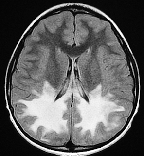
·
T2/FLAIR hyperintensities in the
periventricular and deep white matter
·
Involvement of the corpus callosum, internal capsule, and centrum semiovale
·
Possible sparing of U-fibers (in certain
subtypes)
·
Symmetrical pattern typical of metabolic
disorders
Diagnosis
MRI is the gold standard for detecting
leukodystrophies. CT may help assess chronic atrophy or calcifications.
Definitive diagnosis requires:
·
Clinical correlation
·
Genetic testing
·
Enzyme assays
(e.g., arylsulfatase A for MLD)
·
Nerve conduction studies
Treatment
Current treatments are largely
supportive:
·
Physical and occupational therapy
·
Anticonvulsants for seizure management
·
Baclofen for spasticity
·
Feeding and communication aids
Experimental Therapies:
·
Gene therapy
(e.g., lentiviral vector for ALD)
·
Hematopoietic stem cell transplantation
(HSCT)
·
Enzyme replacement therapy (ERT)
(ongoing trials)
Prognosis
Prognosis varies:
·
Infantile forms often lead to death
within the first decade
·
Late-onset cases may have slower
progression
·
Early diagnosis and therapy initiation
may improve outcomes, especially in ALD and MLD with pre-symptomatic HSCT
Quiz
1. Which imaging modality is most useful
for diagnosing leukodystrophy?
A. CT
B. MRI
C. PET
D. Ultrasound
Explanation: MRI is more sensitive for detecting
white matter changes and characterizing the pattern of demyelination.
2. What is a common clinical feature of
adult-onset leukodystrophy?
A. Fever
B. Acute
hemiplegia
C. Progressive cognitive decline
D. Sudden vision
loss
Explanation: Adult-onset leukodystrophy often
presents insidiously with cognitive impairment and behavioral changes.
3. What therapeutic option is being
explored for certain leukodystrophies like ALD?
A. Radiation
therapy
B.
Corticosteroids
C. Gene therapy
D. Surgical
decompression
Explanation: Gene therapy using lentiviral vectors
has shown promise in treating childhood cerebral ALD.
Conclusion
Leukodystrophy is a complex, devastating
condition requiring high clinical suspicion and advanced imaging for accurate
diagnosis. Despite limited treatments, ongoing gene and stem cell therapies
offer hope. Increased awareness and early detection are key to improving
prognosis.
References
1.
van der Knaap MS, Bugiani M.
Leukodystrophies: a proposed classification system based on pathological
changes and pathogenetic mechanisms. Acta
Neuropathol. 2017;134(3):351–382. doi: 10.1007/s00401-017-1719-x
2.
Vanderver A, Prust M, Tonduti D, et al.
Case definition and classification of leukodystrophies and
leukoencephalopathies. Mol Genet Metab.
2015;114(4):494–500. doi: 10.1016/j.ymgme.2015.01.008
3.
Eichler F, Duncan C, Musolino PL, et al.
Gene therapy for adrenoleukodystrophy. N Engl
J Med. 2017;377(17):1630–1638. doi: 10.1056/NEJMoa1700554
4.
Pouwels PJ, Vanderver A, Bernard G, et
al. Hypomyelinating leukodystrophies: translational research and future
therapies. Neurology.
2014;83(5):463–470. doi: 10.1212/WNL.0000000000000643
5.
Waldman A, Tzarouchi LC, Tambasco N, et
al. Adult-onset leukodystrophies: a clinical and radiological challenge. J Neurol Neurosurg Psychiatry.
2020;91(4):448–456. doi: 10.1136/jnnp-2019-321075
6.
van Rappard DF, Boelens JJ, Wolf NI.
Metachromatic leukodystrophy: disease spectrum and approaches for treatment. Best Pract Res Clin Endocrinol Metab.
2015;29(2):261–273. doi: 10.1016/j.beem.2014.10.002
7. Saudubray JM, Garcia-Cazorla A. Inborn Errors of Metabolism Overview: Pathophysiology, Manifestations, and Management. Pediatr Clin North Am. 2018;65(2):179–208. doi: 10.1016/j.pcl.2017.11.001
leukodystrophy, adult-onset leukodystrophy, white matter disease, brain MRI, leukodystrophy CT, metachromatic leukodystrophy, adrenoleukodystrophy, myelin disorders, neurometabolic diseases, brain atrophy MRI, gene therapy for leukodystrophy, rare brain disorders, white matter MRI abnormalities, leukodystrophy treatment, leukodystrophy imaging


Comments
Post a Comment