Tuberculous Cervical Lymphadenitis: The Silent Swelling in the Neck
Tuberculous Cervical Lymphadenitis: The Silent Swelling in the Neck
Author: Dr. Elias Lee
Category: Infectious Disease | Radiology | Tuberculosis
Estimated reading time: 5 minutes
Introduction
Tuberculous cervical
lymphadenitis, also known as scrofula, remains the most common form of
extrapulmonary tuberculosis (TB), particularly in developing countries and
immunocompromised individuals. In this column, we explore a textbook case of a
22-year-old female presenting with bilateral neck masses, whose CT findings
reveal the classic radiologic signs of this disease.
Clinical Case Summary
A 22-year-old previously
healthy woman presented with progressive, painless swelling on both sides of
the neck and difficulty swallowing. She denied systemic symptoms such as fever,
sore throat, and shortness of breath.
Neck CT with contrast showed:
- Massively enlarged necrotic lymph nodes at levels II
and III bilaterally
- Thick rim enhancement suggestive of central necrosis
- No mass in the salivary glands or aerodigestive
tract
- Prominent lymphoid tissue in the nasopharynx and
palatine tonsils

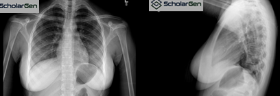
Chest X-ray was unremarkable, showing no
signs of active pulmonary TB.
Final Diagnosis: Tuberculous cervical
lymphadenitis
Pathophysiology &
Epidemiology
Extrapulmonary TB accounts for
~20% of all TB cases in the United States, with lymphadenitis being the most
common form. Cervical involvement is frequently due to hematogenous
reactivation of latent TB bacilli. Risk groups include:
- HIV/AIDS patients
- Immunosuppressed individuals
- Immigrants from TB-endemic regions
Imaging Pearls
Contrast-enhanced CT features
include:
- Rim-enhancing necrotic nodes
- Potential calcifications
- Soft tissue inflammatory changes
- Normal thyroid and salivary glands
Chest imaging is essential to rule out concomitant
pulmonary TB.
Treatment
Standard RIPE therapy includes:
- Rifampin
- Isoniazid
- Pyrazinamide
- Ethambutol
Duration: Minimum 6 months depending on drug susceptibility.
Quiz
1. What is the most likely abnormality
seen on the contrast-enhanced neck CT?
A)
Hypoattenuating lesions in the bilateral tonsils
B) Posterior nasopharyngeal mass
C) Bilateral masses centered in the carotid bodies
with flow voids
D) Bilateral
cystic/necrotic cervical lymphadenopathy
E) Lymphoepithelial cysts in the bilateral parotid
glands
Explanation: This is a classic
finding in tuberculous lymphadenitis. The CT images demonstrate enlarged level
II and III lymph nodes with central low attenuation and rim enhancement,
indicating necrosis. These features are hallmarks of TB lymphadenitis.
2. What
is the most appropriate next step in clinical management?
A)
Place the patient in airborne isolation
B) Direct tissue sampling
C) Sputum sampling
D) HIV testing
E) Further imaging
Explanation: The next critical step
is to confirm the diagnosis via fine-needle aspiration (FNA) or excisional
biopsy. Microbiologic testing (e.g., acid-fast bacilli smear, PCR, culture) is
essential for TB confirmation and drug susceptibility testing. Airborne
isolation or HIV testing may be warranted but are not the immediate next steps.
3. A
workup for mycobacterial infection should be obtained.
A) TRUE
B) FALSE
Explanation: Given the imaging and
clinical context of bilateral necrotic lymphadenopathy, mycobacterial infection
(especially TB) must be high on the differential list. Appropriate workup
includes TB PCR, culture, and histopathology.
4. Which
imaging study should be performed next?
A)
CT head
B) CT angiogram of the neck
C) Chest x-ray
D) Full-body PET/CT
Explanation: Even in cases where
cervical TB is suspected, it is critical to rule out pulmonary involvement,
which determines infection control precautions and guides treatment. Chest
radiography is fast, accessible, and highly informative.
5. The
patient has a normal chest x-ray.
A) TRUE
B) FALSE
Explanation: The chest X-ray
revealed no active disease, pleural effusion, or lung involvement. However, it
is important to note that normal chest X-ray findings do not exclude TB
lymphadenitis, as up to 50% of these cases occur without pulmonary disease.
6. Which
of the following is NOT part of the first-line treatment for TB lymphadenitis?
A)
Rifampin
B) Isoniazid
C) Pyrazinamide
D) Clarithromycin
E) Ethambutol
Explanation: The first-line RIPE
regimen for TB includes Rifampin, Isoniazid, Pyrazinamide, and Ethambutol.
Clarithromycin is used for nontuberculous mycobacterial infections, not Mycobacterium tuberculosis.
References
- Alvarez S, McCabe WR. Extrapulmonary tuberculosis
revisited: a review of experience at Boston City and other hospitals.
Medicine (Baltimore). 1984;63(1):25–55.
- Moon WK, Han MH, Chang KH, et al. CT and MR
imaging of head and neck tuberculosis. Radiographics.
1997;17(2):391–402.
- Peto HM, et al. Epidemiology of extrapulmonary
tuberculosis in the United States, 1993–2006. Clin Infect Dis.
2009;49(9):1350–1357.
- Rieder HL, Snider DE Jr, Cauthen GM. Extrapulmonary
tuberculosis in the United States. Am Rev Respir Dis.
1990;141(2):347–351.
- Fontanilla JM, Barnes A, von Reyn CF. Current
diagnosis and management of peripheral tuberculous lymphadenitis. Clin
Infect Dis. 2011;53(6):555–562.
- Lee Y, et al. Ultrasonography and CT findings of
tuberculous lymphadenitis. Korean J Radiol. 2001;2(4):210–215.
- World Health Organization. Global tuberculosis
report 2023. Geneva: WHO; 2023.


Comments
Post a Comment