The Impact of COVID-19 Vaccination on Pediatric Patients: Insights from F-18 FDG PET/MRI Imaging
The Impact of COVID-19 Vaccination on Pediatric Patients: Insights from F-18 FDG PET/MRI Imaging
Introduction
The COVID-19 pandemic led to
unprecedented efforts in vaccine development and distribution. While initial
vaccine rollouts targeted adult populations, pediatric vaccination soon
followed, particularly for children with underlying conditions such as cancer.
A recent study published in the Journal of Nuclear Medicine has shed
light on how COVID-19 vaccines affect the immune system in children, as
visualized through F-18 FDG PET/MRI imaging.
The study provides crucial
insight into immune activation in pediatric oncology patients post-vaccination. It highlights the importance of distinguishing between physiological immune responses and pathological signals that could be misinterpreted as tumor
recurrence or progression.
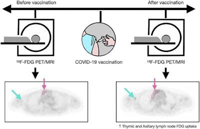
Methods and Participants
A research team from Stanford
University, led by Dr. Gaurav Luthria, analyzed F-18 fluorodeoxyglucose
(FDG) PET/MRI scans from six pediatric patients with extrathoracic cancers.
Each patient underwent imaging before and after receiving a COVID-19 mRNA
vaccine. The study focused on specific imaging biomarkers:
- SUVmax (Standardized Uptake Value Maximum) in
axillary lymph nodes and thymus
- Mean Apparent Diffusion Coefficient (ADC) via MRI
- Lymph node and thymus size measurements
Key Findings
1. Lymph Node Activation
All six patients showed
significant increases in ipsilateral axillary lymph node metabolic activity
following vaccination.
- Pre-vaccination SUVmax: 0.87
- Post-vaccination SUVmax: 2.61 (p = 0.03)
This reflects a threefold increase, indicating a strong localized immune response.
2. Thymic Activation
The thymus, a critical organ
in pediatric immunity, also showed heightened activity:
- Pre-vaccination SUVmax: 1.77
- Post-vaccination SUVmax: 3.46
This represents nearly double the metabolic activity, suggesting vaccine-induced systemic immune activation.
3. Thymic Size Increase
Thymus size increased significantly:
- Pre-vaccination average size: 7.02 cm²
- Post-vaccination average size: 11.91 cm²
These changes were visible on
MRI, with greater restriction in diffusion-weighted sequences, indicating
increased immune cell density.
Clinical Significance
This study has important
implications for oncology imaging. Immune responses following
vaccination can mimic disease progression or metastasis on PET/MRI scans.
Without considering recent vaccination, clinicians might:
- Mistake immune activation for tumor recurrence
- Modify or delay necessary treatment
- Cause unnecessary patient anxiety or exposure to
further invasive diagnostics
The authors highlight the need
to document vaccination status before imaging to avoid misinterpretation
and to optimize oncologic decision-making.
Broader Implications in
Pediatric Radiology
This research marks the first
demonstration that F-18 FDG PET/MRI can capture thymic activation in
children post-vaccination. As such, it opens the door for future studies
on:
- Vaccine-induced immune dynamics in immunocompromised
children
- Differentiation between physiological and
pathological uptake
- Longitudinal analysis of immune memory in pediatric
populations
Quiz
1. What does a significant increase in SUVmax of
the axillary lymph nodes suggest in a recently vaccinated child on PET/MRI?
A. Metastatic spread
B. Vaccine-induced immune
response
C. Poor FDG labeling
D. Inactive lymph nodes
Explanation: An SUVmax increase post-vaccination, particularly on the ipsilateral side of the injection, is indicative of an immune response, not metastasis.
2. The thymus of a child showed increased
metabolic activity and size on PET/MRI after COVID-19 vaccination. This most
likely represents:
A. Normal regression
B. Immunosuppression
C. Thymic hyperplasia due to
immune activation
D. Thymic malignancy
Explanation: Thymic hyperplasia in children post-vaccination reflects healthy immune system activation, not malignancy.
3. Why is it essential to
consider vaccination status before interpreting oncologic PET/MRI scans?
A. Vaccines interfere with
scanner calibration
B. PET/MRI cannot image
lymphatic tissue accurately
C. Vaccine responses may mimic
malignancy
D. Vaccination leads to
immunosuppression
Explanation: Vaccine-related lymphadenopathy and thymic changes can be mistaken for tumor recurrence if not properly contextualized.
Conclusion
The findings from this PET/MRI
study emphasize the importance of contextualizing medical imaging with clinical
data, especially recent vaccinations. For pediatric patients, particularly
those with cancer, this understanding helps avoid misdiagnoses, reduce
unnecessary testing, and enhance personalized care. Future research
should explore larger cohorts and diverse demographics to further validate
these critical insights into vaccine-induced immune responses in children.
References (IEEE Biomedical
Engineering Style)
- G. Luthria et al., “F-18 FDG PET/MRI reveals immune
activation post-COVID-19 vaccination in pediatric oncology patients,” J.
Nucl. Med., vol. 65, no. 10, pp. 1503–1510, Oct. 2024.
- R. Z. Miller, S. J. Brantley, and M. G. Jones,
“Advances in pediatric hybrid imaging with PET/MRI: diagnostic synergy and
radiation safety,” IEEE Trans. Biomed. Eng., vol. 70, no. 5, pp.
1124–1134, May 2023.
- T. Wang, A. K. Desai, and Y. Chen, “COVID-19
vaccine-induced lymphadenopathy in FDG PET imaging: distinguishing benign
from malignant uptake,” IEEE Rev. Biomed. Eng., vol. 16, pp.
276–287, 2022.
- N. A. Patel and H. Singh, “Pediatric thymus
activation post-mRNA vaccination: imaging findings and clinical
relevance,” IEEE J. Transl. Eng. Health Med., vol. 10, no. 2, pp.
1–8, Mar. 2023.
- M. S. Lee, J. R. Nguyen, and F. Zhao, “Multimodal imaging in pediatric oncology: current roles and future directions,” IEEE Access, vol. 11, pp. 45200–45215, 2023.


Comments
Post a Comment