Osteonecrosis of the hip
Osteonecrosis of the hip
Osteonecrosis of the hip (also called avascular necrosis [AVN] of the
femoral head) is a progressive, debilitating condition that results from interrupted
blood supply to the femoral head, leading to bone ischemia, necrosis,
and collapse of the articular surface.
1. Cause and Etiology
Osteonecrosis can be traumatic or non-traumatic:
A. Traumatic Osteonecrosis
- Cause: Disruption of blood supply due to:
- Femoral neck fractures
- Hip dislocations
- Iatrogenic injury (e.g., after hip
surgery)
- Trauma disrupts retinacular arteries,
branches of the medial femoral circumflex artery, leading to
ischemia.
B. Non-Traumatic Osteonecrosis
- Multifactorial, usually involving vascular
compromise and cellular apoptosis.
- Major risk factors include:
- Corticosteroid use (the most common
non-traumatic cause)
- Excessive alcohol intake
- Sickle cell disease
- Systemic lupus erythematosus
- Organ transplantation
- Decompression sickness (caisson disease)
- Hypercoagulable states (e.g., Factor V Leiden)
- Autoimmune diseases
- HIV infection
- Pancreatitis
- Radiation therapy
- Idiopathic (~20–40% cases)
2. Pathophysiology
The disease progresses through the following pathophysiological stages:
- Ischemia of the femoral head due
to interruption of blood flow.
- Death of osteocytes and bone marrow components within 24–72 hours.
- Repair attempts begin at the periphery,
but necrotic bone cannot be resorbed and replaced fast enough.
- Subchondral collapse occurs due to weakened
bone beneath the cartilage.
- Articular cartilage failure and joint incongruity
lead to secondary osteoarthritis.
3. Epidemiology
- Affects young to middle-aged adults, usually
between 30–50 years.
- Bilateral involvement occurs in 30–70%
of cases.
- Males > females (about 2:1 ratio).
- The incidence is difficult to determine, but
estimates in the U.S. suggest 10,000–20,000 new cases/year.
- Up to 50% of nontraumatic osteonecrosis cases
are related to corticosteroid use or alcohol.
4. Clinical Presentation
A. Symptoms
- Groin pain is the most common
initial symptom.
- Pain can radiate to the buttock, thigh,
or knee.
- Pain worsens with weight-bearing or hip
movement.
- Range of motion becomes progressively
limited.
- Limping may develop.
B. Progression
- Initially asymptomatic (early stages).
- Pain increases as subchondral collapse
develops.
- Advanced stages lead to degenerative arthritis.
5. Imaging Features
A. Plain Radiographs

- Normal in early stages.
- Late findings:
- Crescent sign: Subchondral
radiolucency (subchondral fracture).
- Femoral head flattening and joint space
narrowing.
- Secondary osteoarthritis.
B. MRI (Most Sensitive
Modality)

- Detects early changes before the X-ray is abnormal.
- Findings:
- Double line sign: Inner bright T2 line
(granulation tissue) + outer dark line (sclerosis).
- Geographic low signal on T1.
- Bone marrow edema in acute phases.
C. CT Scan

- Useful for detecting subchondral fracture and
sclerosis.
- Helps in preoperative planning.
D. Bone Scintigraphy

- Increased uptake in early stages.
- Less specific than MRI.
6. Treatment
Treatment depends on stage, extent of involvement, and patient
symptoms.
A. Non-Surgical (Early Stage)
- Activity modification and limited weight
bearing.
- Pharmacologic agents:
- Bisphosphonates (e.g., alendronate): Delay
collapse.
- Statins: Theoretical role in lipid metabolism.
- Anticoagulants: If thrombophilia is present.
- Vasodilators (e.g., iloprost).
- Electrical stimulation or shockwave therapy (experimental).
B. Surgical Management
- Core Decompression:
- Drilling into the necrotic area to reduce
intraosseous pressure.
- Effective in early disease (Ficat I-II).
- Can be combined with bone grafting
(vascularized or non-vascularized).
- Osteotomy:
- Realigns the joint to shift weight-bearing away
from the necrotic region.
- Less commonly used.
- Bone Grafting:
- Vascularized fibular grafts: Restore blood supply
and structural support.
- Total Hip Arthroplasty (THA):
- Indicated for advanced disease with joint
collapse and arthritis.
- Excellent pain relief and function restoration.
7. Prognosis
- Highly dependent on early diagnosis and the extent
of femoral head involvement.
- Without treatment:
- 80% of patients progress to collapse
and secondary arthritis.
- Smaller, asymptomatic lesions may remain stable.
- Early intervention (e.g., core
decompression) may preserve the joint.
- Total hip replacement has excellent long-term
outcomes but is not ideal for younger patients due to implant lifespan and the need for revision.
8. Classification Systems
A. Ficat and Arlet
Classification:
- Stage 0: Normal imaging, histologic changes only.
- Stage I: Normal X-ray, MRI positive.
- Stage II: Sclerosis/cyst
formation, no collapse.
- Stage III: Subchondral collapse
(crescent sign), but joint space preserved.
- Stage IV: Joint space narrowing,
secondary osteoarthritis.
Case study: A 38-Year-Old Female with Chronic Hip Pain
Osteonecrosis of the Hip
Clinical History and Imaging Findings
-
A 38-year-old female presented with a chief complaint of chronic pain in the left hip.
-
The pain had progressively worsened following a fall two weeks before presentation. The patient has a history of being a professional athlete.
-
Radiographic imaging of the hip was obtained for further evaluation.

Quiz 1
-
What does the red arrow indicate?
(1) A crescent-shaped subchondral lucency, suggestive of a subchondral fracture
(2) Subchondral cyst formation due to osteoarthritis
(3) Joint space narrowing
(4) Marginal erosion due to inflammatory changesExplanation:
The red arrow points to a crescent-shaped subchondral lucency, consistent with a subchondral fracture. Subchondral cysts and marginal erosions are more commonly associated with inflammatory arthropathies, such as rheumatoid arthritis. Although there are radiographic features suggestive of osteoarthritis in this patient, the red arrow specifically highlights a subchondral fracture indicative of articular surface collapse. -
Which imaging modality would you recommend next?
(1) CT
(2) MRI
(3) Bone scintigraphyExplanation:
While bone scintigraphy can be useful in differentiating bone marrow edema, insufficiency fractures, and osteonecrosis, MRI provides the most definitive diagnostic information. It is also invaluable for surgical planning.
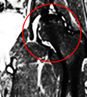
MRI Findings
An MRI of the hip was performed for further evaluation.

Postoperative Follow-Up
The patient underwent total hip arthroplasty. A follow-up pelvic radiograph was obtained for postoperative evaluation.

Quiz 2
-
What is the most likely diagnosis?
(1) Transient bone marrow edema
(2) Rheumatoid arthritis
(3) Osteonecrosis
(4) Acute insufficiency fractureExplanation:
The imaging features are most consistent with osteonecrosis (ON) of the hip. While ON can occur in the setting of rheumatoid arthritis, such cases are typically accompanied by additional inflammatory findings. In transient bone marrow edema syndrome (also known as transient osteoporosis of the hip), the crescent-shaped signal alteration typically seen in ON is absent. Although insufficiency fractures may also present with a low-signal intensity band, it tends to be convex toward the articular surface, as opposed to the concave configuration often seen in ON. -
Which of the following is the most characteristic or pathognomonic MRI finding in this case?
(1) Bone marrow edema
(2) Double-line sign
(3) Linear sclerosis due to pathological fracture
(4) Synovial hypertrophy with pannus formationExplanation:
The double-line sign is a highly characteristic and specific MRI finding for osteonecrosis of the hip. It consists of a serpiginous low-signal-intensity outer rim representing sclerotic bone, and a high-signal-intensity inner line corresponding to granulation tissue. As demonstrated in this case, the double-line sign is a hallmark feature of ON on T2-weighted or STIR sequences.

3. What is the pathogenesis of pain in osteonecrosis (ON)?
(1) Increased intraosseous pressure from bone marrow edema causes pain
(2) Inflammation affects the periosteum
(3) Bone death and necrosis directly cause pain
Explanation:
Osteonecrosis (ON) is typically asymptomatic in its early stages. When pain does develop, it is often due to increased intraosseous pressure resulting from bone marrow edema. The long-term prognosis is primarily determined by whether the articular surface of the femoral head collapses. Therefore, assessing for evidence of subchondral collapse is critical.
4. Osteonecrosis of the hip is frequently bilateral and often asymptomatic in the early stages. Therefore, it is important to image both hips.
(1) True
(2) False
Explanation:
Up to 38% of patients with osteonecrosis of the hip may be asymptomatic. While traumatic osteonecrosis is typically unilateral, systemic causes such as corticosteroid use, excessive alcohol intake, sickle cell anemia, or idiopathic etiologies are often bilateral. Consequently, both hips should be thoroughly evaluated, even if symptoms are unilateral.
Findings and Diagnosis
Preoperative Radiographs:

There is bilateral avascular necrosis (osteonecrosis) of the femoral heads. The left femoral head demonstrates flattening with associated subchondral collapse of the articular surface. Superimposed mild secondary osteoarthritis is present bilaterally, characterized by joint space narrowing and a large subchondral cyst in the superior aspect of the left acetabulum. Additionally, an intra-articular osteochondral loose body is noted inferior-medial to the left hip joint. The remainder of the pelvic structures appears intact.
Preoperative MRI:

The left hip demonstrates findings consistent with osteonecrosis (ON), including low-signal intensity fibrotic changes and mild flattening of the anterosuperior femoral head, indicative of early subchondral collapse of the articular surface.
There is superimposed secondary osteoarthritis, more pronounced on the left than the right, characterized by marginal spurring and subchondral cystic changes.

Postoperative radiographs were obtained following total hip arthroplasty.

A left total hip arthroplasty prosthesis is in place. Sclerosis of the right femoral head is noted, consistent with avascular necrosis. No evidence of articular surface collapse is observed.
Differential Diagnosis
-
Transient Bone Marrow Edema Syndrome (Transient Osteoporosis of the Hip)
Similar features to ON:-
Osteopenia
-
Bone marrow edema
Key distinguishing features from ON: -
In transient bone marrow edema, the uptake of radioactive isotopes is significantly increased and diffuse, unlike the central hypointensity observed in ON.
-
No crescent-shaped signal changes are seen in transient bone marrow edema.
-
-
Acute Insufficiency Fracture
Similar features to ON:-
Low-signal intensity band
Key distinguishing features from ON: -
The sclerotic band in an insufficiency fracture is typically convex, unlike the concave and serpentine appearance seen in ON.
-
A more extensive bone marrow edema is expected in insufficiency fractures.
-
Diagnosis:
Osteonecrosis of the Hip
Discussion
Osteonecrosis (ON)
Epidemiology:
Osteonecrosis (ON) is a common musculoskeletal disorder, with approximately 10,000 to 20,000 new cases of osteonecrosis reported annually. However, due to the high incidence of asymptomatic cases, this estimate is likely an underrepresentation. The most common causes of femoral head osteonecrosis are trauma, corticosteroids, alcohol use, and idiopathic origins. The prevalence of corticosteroid-induced osteonecrosis ranges from 3% to 38%, with the highest rates reported in patients receiving corticosteroid therapy for acute lymphoblastic leukemia.
Pathophysiology:
ON occurs when the blood supply to the epiphysis or subchondral bone is disrupted, leading to necrosis of the trabecular bone. This results in ischemic bone, which may cause hemorrhage. The reactive interface attempts to isolate the necrotic tissue. Osteoblasts and osteoclasts work to deposit new bone and absorb the damaged tissue, progressing towards subchondral sclerosis. As the articular surface progressively deteriorates, cortical flattening and collapse occur in the later stages.
Clinical Presentation:
ON can often be asymptomatic, but when symptoms do appear, they typically include pain and reduced range of motion. Pain results from increased intraosseous pressure caused by bone marrow edema. Long-term prognosis is largely determined by whether collapse of the femoral head articular surface occurs. Therefore, it is crucial to assess for the presence of superior articular surface collapse.
Imaging Features:
-
X-ray:
Early imaging may show normal radiographs or subtle areas of localized translucency and sclerosis in the femoral head. As lateral cartilage rupture progresses in ON, characteristic serpentine sclerotic borders become evident, usually observed in the diaphyseal region. Articular collapse is generally noted at the junction of the sclerotic border and the articular surface, with a crescent-shaped lucency (crescent sign) in the subchondral area. Secondary osteoarthritis may develop as the lateral cartilage rupture progresses. -
MRI:
Subchondral sclerosis is typically seen on T1-weighted images, most prominently in the anterior and superior femoral head, visible on coronal imaging. The double-line sign, featuring a low-signal intensity outer rim and a high-signal intensity inner area representing granulation tissue, is pathognomonic for osteonecrosis. This double-line sign, as seen in this case, is a key feature of subchondral sclerosis in ON.
MRI Steinberg Classification of Osteonecrosis
| Stage | Description | MRI Findings |
|---|---|---|
| Stage I | Early Stage (Pre-collapse) | Normal MRI or mild changes with subchondral edema and no articular surface collapse. |
| Stage II | Intermediate Stage (Collapse begins) | Subchondral sclerosis and cyst formation. No collapse of the articular surface yet. |
| Stage III | Advanced Stage (Partial Collapse) | Partial collapse of the femoral head with a crescent sign and subchondral cysts. |
| Stage IV | End Stage (Complete Collapse) | Complete collapse of the femoral head, often with extensive secondary osteoarthritis and joint space narrowing. |
Treatment:
Core decompression can help alleviate symptomatic pain by reducing intraosseous pressure. This procedure is suitable for patients without significant subchondral collapse. For patients with advanced secondary osteoarthritis, total hip arthroplasty (THA) is required.
References:
(1) Murphey MD, Foreman KL,
Klassen-Fischer MK, Fox MG, Chung EM, Kransdorf MJ. From the radiologic
pathology archives imaging of osteonecrosis: Radiologic-pathologic correlation.
RadioGraphics. 2014;34(4):1003-1028. doi:10.1148/rg.344140019
(2) Assouline-Dayan Y, Chang C,
Greenspan A, Shoenfeld Y, Gershwin ME. Pathogenesis and natural history of
osteonecrosis. Semin Arthritis Rheum. 2002;32(2):94-124.
(3) Ito H, Matsuno T, Minami A.
Relationship between bone marrow edema and development of symptoms in patients
with osteonecrosis of the femoral head. AJR Am J Roentgenol.
2006;186(6):1761-1770. doi:10.2214/AJR.05.0086
(4) Stevens K, Tao C, Lee SU,
et al. Subchondral fractures in osteonecrosis of the femoral head: Comparison
of radiography, CT, and MR imaging. Am J Roentgenol. 2003;180(2):363-368.
doi:10.2214/ajr.180.2.1800363
(5) Steinberg ME, Hayken GD,
Steinberg DR. A quantitative system for staging avascular necrosis. J Bone
Joint Surg Br. 1995;77:34-41.
(6) Pierce TP, Jauregui JJ,
Elmallah RK, Lavernia CJ, Mont MA, Nace J. A current review of core
decompression in the treatment of osteonecrosis of the femoral head. Curr Rev
Musculoskelet Med. 2015;8(3):228-232. doi:10.1007/s12178-015-9280-0
(7) Steinberg ME, Hayken GD,
Steinberg DR. A quantitative system for staging avascular necrosis. J Bone
Joint Surg Br. 1995 Jan;77(1):34-41. PMID: 7822393


Comments
Post a Comment