Discovery of X-Rays
The Discovery of X-Rays by Wilhelm Conrad Röntgen and Its Enduring Influence on Modern Medicine
Abstract
The accidental discovery of X-rays by Wilhelm Conrad Röntgen in 1895 marked a monumental shift in both the scientific and medical worlds. Röntgen’s work not only earned him the first Nobel Prize in Physics in 1901 but also laid the groundwork for the birth of diagnostic radiology—a field now indispensable to modern medicine. This paper traces the historical development and scientific foundations of X-ray imaging, evaluates its medical applications in diagnostics and therapeutics, and explores contemporary innovations such as computed tomography (CT), digital radiography, and artificial intelligence (AI)-assisted diagnostics. It also discusses ongoing challenges related to radiation safety and considers future trends in imaging technology.
1. Introduction
Prior to the advent of imaging technology, physicians faced substantial limitations in diagnosing and treating internal diseases. Exploratory surgery and physical examination remained the primary methods for investigating pathology, often at considerable risk to patients. That paradigm changed dramatically in late 1895, when Wilhelm Conrad Röntgen discovered a new type of electromagnetic radiation, which he called “X-rays” due to their unknown nature.
Röntgen’s ability to visualize the bones within his wife's hand using this mysterious form of energy stunned the global scientific community. Medical practitioners quickly grasped the implications of this technology, initiating a new era in non-invasive diagnostics. Over time, the development of sophisticated imaging techniques, including CT scans, angiography, and interventional radiology, has dramatically expanded the utility of X-rays in clinical settings. Röntgen’s legacy lives on in every modern hospital, where radiology departments are vital to patient care.
2. The Discovery of X-Rays
2.1 The Scientific Context
The late 19th century was a fertile period for physics, with intense interest in cathode rays and the nature of electromagnetic energy. Wilhelm Röntgen, then a professor of physics at the University of Würzburg, was experimenting with Crookes tubes—devices that emitted cathode rays under high voltage. On November 8, 1895, he observed an unexplained fluorescence on a barium platinocyanide-coated screen positioned a few feet away from the tube, even though the tube was enclosed in black cardboard.
This observation led him to conduct a series of methodical experiments. He discovered that the rays passed through most substances, but not dense materials like bone or lead. He coined the term “X-rays” to signify their mysterious qualities. Within weeks, Röntgen captured the now-famous image of his wife Bertha's hand, which showed her skeletal structure and wedding ring (Glasser, 1993).

2.2 Early Medical Applications
The medical utility of X-rays was quickly realized. By 1896, hospitals in Europe and North America had adopted primitive X-ray equipment. During the Greco-Turkish War (1897), battlefield medics used X-rays to locate bullets in wounded soldiers. However, the dangers of prolonged exposure to ionizing radiation were not yet understood. Early radiologists often suffered from severe burns, radiation-induced cancers, and even amputations (Bushberg et al., 2011).
Despite these risks, the benefits of X-ray imaging outweighed the drawbacks, and it became increasingly integral to medical diagnostics.
3. The Role of X-Rays in Modern Medical Practice
3.1 Diagnostic Radiology
Today, X-ray imaging is one of the most commonly used diagnostic tools worldwide. It is essential for evaluating musculoskeletal injuries, pulmonary diseases, dental conditions, and numerous other pathologies. The high spatial resolution and rapid image acquisition make conventional radiography invaluable in emergency departments and outpatient clinics.
Contrast-enhanced X-ray techniques, such as barium swallow studies and intravenous pyelography, have broadened the range of observable structures, including the gastrointestinal and urinary tracts. Fluoroscopy, the real-time application of X-ray imaging, is extensively used for dynamic evaluations such as swallowing studies and cardiac catheterization procedures (Bushberg et al., 2011).
3.2 Computed Tomography (CT)
One of the most significant advances derived from X-ray technology is computed tomography. Developed by Godfrey Hounsfield and Allan Cormack in the early 1970s, CT allows cross-sectional imaging of the body, offering unprecedented detail. By rotating an X-ray source and detector around the patient, a computer constructs three-dimensional images of tissues and organs.
CT scans are instrumental in diagnosing cerebrovascular accidents, neoplasms, traumatic injuries, and infections. Dual-energy CT and contrast-enhanced protocols further enhance diagnostic precision (Kalra et al., 2004).
3.3 Interventional Radiology
X-ray fluoroscopy forms the backbone of interventional radiology—a specialty that performs minimally invasive procedures under image guidance. Applications range from angioplasty and embolization to vertebroplasty and tumor ablation. These procedures offer reduced morbidity and quicker recovery times compared to traditional surgery (Hall & Giaccia, 2012).
3.4 Radiation Therapy
X-rays are also employed therapeutically in radiation oncology. High-energy X-rays can destroy or damage cancer cells, and modern techniques such as intensity-modulated radiation therapy (IMRT) and stereotactic body radiotherapy (SBRT) allow for precise targeting of tumors while sparing healthy tissue.
4. Technological Innovations and Contemporary Issues
4.1 Digital Radiography
The shift from analog film to digital radiography has significantly improved workflow, image quality, and data storage. Digital systems allow real-time image processing, enhanced contrast resolution, and seamless integration with hospital information systems. They also support teleradiology, enabling remote diagnosis and consultation (Oakden-Rayner, 2017).
4.2 Artificial Intelligence in Imaging
AI applications in radiology are advancing rapidly. Deep learning algorithms are being trained to detect anomalies such as fractures, nodules, and hemorrhages with high accuracy. AI enhances efficiency by prioritizing urgent cases, reducing radiologist fatigue, and supporting clinical decision-making (Oakden-Rayner, 2017).

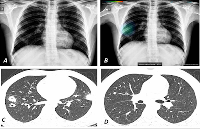
This chest radiograph demonstrates how AI-based software automatically detects pulmonary lesions and highlights the affected areas using a color overlay.
|
The color overlay visually represents both the location and severity of the lesions, helping clinicians identify abnormalities quickly and accurately. Such AI-assisted diagnostic tools are particularly useful for detecting early-stage lung nodules and other subtle lesions that might otherwise be overlooked. |
4.3 Radiation Safety
A persistent concern in X-ray imaging is radiation exposure. While the doses are generally low for standard radiography, repeated exposure, especially from CT scans, can accumulate. Pediatric imaging is particularly sensitive. Techniques such as dose modulation, shielding, and judicious use of imaging are critical to minimize harm (Brenner & Hall, 2007).
4.4 The Future of X-Ray Imaging
Emerging technologies include spectral (dual-energy) CT and photon-counting detectors, which promise greater tissue contrast and lower radiation doses. In tandem with AI and molecular imaging techniques, the future of X-ray imaging will likely involve hybrid modalities and personalized diagnostics.
5. Conclusion
Wilhelm Conrad Röntgen’s discovery of X-rays stands as one of the most significant achievements in the history of science and medicine. It transformed diagnostics, enabled precise therapies, and laid the foundation for the multi-billion-dollar radiology industry. As technology continues to evolve, the basic principles Röntgen uncovered more than a century ago remain at the heart of modern medical imaging.
From its serendipitous origin to its indispensable role in contemporary healthcare, the legacy of X-ray imaging exemplifies the power of scientific discovery to advance human well-being. Looking forward, the integration of artificial intelligence, radiation safety enhancements, and next-generation imaging technologies will ensure that Röntgen’s contribution continues to save lives well into the future.
References
-
Brenner, D. J., & Hall, E. J. (2007). Computed tomography—An increasing source of radiation exposure. New England Journal of Medicine, 357(22), 2277-2284. https://doi.org/10.1056/NEJMra072149
-
Bushberg, J. T., Seibert, J. A., Leidholdt Jr, E. M., & Boone, J. M. (2011). The essential physics of medical imaging (3rd ed.). Lippincott Williams & Wilkins.
-
Glasser, O. (1993). Wilhelm Conrad Röntgen and the early history of the Roentgen rays. Norman Publishing.
-
Hall, E. J., & Giaccia, A. J. (2012). Radiobiology for the radiologist (7th ed.). Lippincott Williams & Wilkins.
-
Kalra, M. K., Maher, M. M., Toth, T. L., Hamberg, L. M., Blake, M. A., Shepard, J. A., & Saini, S. (2004). Strategies for CT radiation dose optimization. Radiology, 230(3), 619–628. https://doi.org/10.1148/radiol.2303031609
-
Oakden-Rayner, L. (2017). Exploring large-scale public medical image datasets. Academic Radiology, 24(6), 744–748. https://doi.org/10.1016/j.acra.2016.10.014


Comments
Post a Comment