Pneumoperitoneum
Pneumoperitoneum
Pneumoperitoneum: Overview
Pneumoperitoneum refers to the presence of
gas or air in the peritoneal cavity, typically indicating a perforation
of a hollow abdominal viscus, which constitutes a surgical emergency.
However, it can also occur in non-emergent settings (non-surgical or spontaneous
pneumoperitoneum).
1. Cause and Etiology
A. Surgical (True)
Pneumoperitoneum – ~90% of cases
Most commonly due to
perforation of an intra-abdominal hollow organ:
- Peptic ulcer disease (gastric or duodenal
perforation)
- Diverticulitis (perforated
diverticulum)
- Appendicitis
- Inflammatory bowel disease (IBD) (e.g., Crohn’s disease
with perforation)
- Bowel obstruction with necrosis/perforation
- Iatrogenic causes
- Laparoscopy (insufflation of CO₂)
- Endoscopy or colonoscopy with perforation
- Peritoneal dialysis catheter insertion
B. Non-surgical (Benign)
Pneumoperitoneum
- Postoperative residual air (resolves within 3–6
days after surgery)
- Mechanical ventilation (barotrauma in
critically ill patients)
- Peritoneal dialysis
- Pneumatosis cystoides intestinalis (gas cysts in bowel wall
rupture)
- Vaginal insufflation (rare, through fallopian
tubes)
- Infections with gas-producing organisms (e.g., Clostridium)
2. Pathophysiology
- In normal conditions, the peritoneal cavity is a
potential space with no air.
- When a viscus perforates, intraluminal gas
(and often contents) escape into the sterile peritoneal cavity.
- This results in chemical and bacterial
peritonitis, leading to:
- Inflammatory response
- Fluid shifts (third spacing)
- Systemic inflammatory response syndrome (SIRS)
- Sepsis or septic shock if untreated
- In benign or non-surgical pneumoperitoneum, there is
no perforation or spillage, so inflammation is minimal or absent.
3. Epidemiology
- Incidence:
- Not precisely quantified due to its presentation as
a radiological sign rather than a disease entity.
- Demographics:
- Affects all age groups.
- Most common in elderly patients due to
higher rates of peptic ulcer disease, diverticulitis, and malignancy.
- Surgical pneumoperitoneum is more common than
spontaneous.
4. Clinical Presentation
A. Perforation-Related
Pneumoperitoneum (Surgical)
- Acute, severe abdominal pain – sudden onset
- Peritonitis signs: guarding, rigidity,
rebound tenderness
- Abdominal distension
- Nausea, vomiting
- Fever and tachycardia
- Hypotension, septic shock (advanced cases)
B. Non-Surgical
Pneumoperitoneum
- Often asymptomatic
- Incidental finding on imaging
- May have mild abdominal discomfort or
bloating
5. Imaging Features
A. Plain Radiograph (X-ray)

- Upright chest X-ray is most sensitive:
- Free air under the diaphragm (especially the right side)
- Supine abdominal X-ray:
- Rigler's sign: visible outlines of
both sides of the bowel wall (air inside and outside)
- Football sign: massive free air
outlining the peritoneal cavity
- Falciform ligament sign: air outlines the falciform
ligament
- Double wall sign: air on both sides of the bowel wall
B. Computed Tomography (CT)

- Gold standard for diagnosis
- Can detect very small amounts of free air
- Can localize the source of perforation
- Shows associated findings: abscess, fluid,
inflammation
C. Ultrasound

- Not first-line but may show reverberation
artifacts (ring-down or comet tail) if free air is present anterior to
the liver.
6. Treatment
A. Surgical Pneumoperitoneum
- Emergency surgery is often required:
- Exploratory laparotomy or laparoscopy to identify and repair the perforation
- Broad-spectrum IV antibiotics
- IV fluid resuscitation
- Nasogastric decompression if ileus or obstruction is present
B. Non-Surgical
Pneumoperitoneum
- Conservative management if no signs of
peritonitis or systemic toxicity
- Close clinical monitoring
- Serial imaging
- Supportive care (e.g., oxygen, fluids)
7. Prognosis
Surgical Pneumoperitoneum
- Prognosis depends on:
- Cause of perforation
- Time to intervention
- Extent of contamination and peritonitis
- Patient comorbidities
- Early intervention can significantly improve
outcomes
- Mortality rates:
- <10% if promptly treated
o
50% if delayed diagnosis or in septic shock
Non-Surgical Pneumoperitoneum
- Excellent prognosis with resolution once the underlying
cause is managed
- Rarely requires surgery
Key Points Summary
|
Aspect |
Summary |
|
Cause |
Mostly due to GI perforation
(e.g., ulcers, diverticulitis); can be non-surgical |
|
Etiology |
Trauma, surgery,
inflammation, infection, and medical procedures |
|
Pathophysiology |
Gas in the peritoneum causes
peritonitis in perforation; benign in others |
|
Epidemiology |
More common in the elderly;
often seen postoperatively or in the ICU |
|
Clinical Features |
Acute abdomen in
perforation; asymptomatic in benign cases |
|
Imaging |
Upright chest X-ray, CT for
confirmation/localization |
|
Treatment |
Surgery for perforation;
observation for benign causes |
|
Prognosis |
Depends on the etiology and the timeliness of treatment |
===================================================
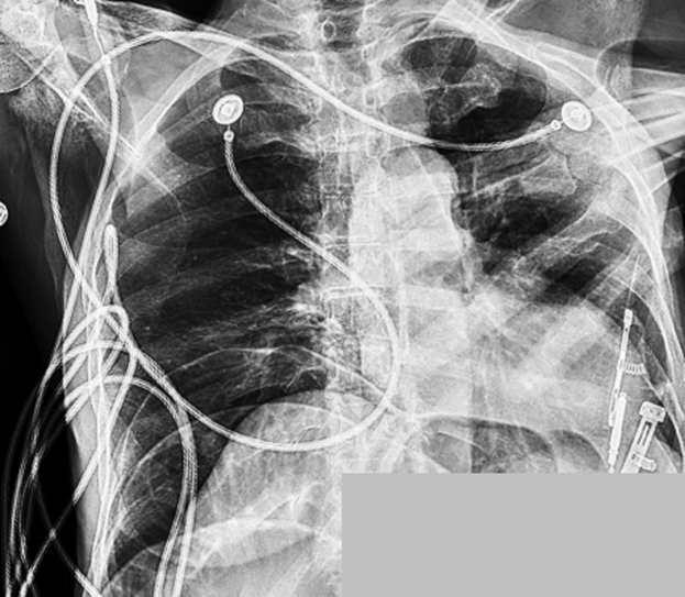
Case Study 792: Altered Mental Status in a 72-Year-Old Female
Pneumoperitoneum
History and Imaging Findings
-
A 72-year-old female presented with altered mental status.
-
Physical examination revealed significant abdominal rigidity and rebound tenderness.
-
Arterial blood gas analysis demonstrated an elevated lactate level of 3.8 mmol/L (reference range: 0.5–1.6 mmol/L).
-
Chart review showed no recent history of surgical procedures.
-
The image below is a portable chest radiograph.
Quiz 1:
-
What is the primary finding on the chest radiograph?
(1) Pneumoperitoneum
(2) Pneumothorax
(3) Pneumomediastinum
(4) Right rib fracture -
There are no benign causes of pneumoperitoneum.
(1) True
(2) False
Explanation:
Recent laparoscopic surgery involving insufflation of the abdomen with carbon dioxide is a benign and non-emergent cause of pneumoperitoneum. Post-traumatic pseudo-pneumoperitoneum is another example of a benign etiology.
Additional Imaging:
-
An emergency contrast-free CT scan of the abdomen and pelvis was obtained (no oral or intravenous contrast was administered).
-
Axial, sagittal, and coronal reformatted images are presented in lung window (first image) and soft tissue window (second and third images).

Quiz 2
-
Which CT findings are indicative of pneumoperitoneum?
A. Outlining of the falciform ligament by free air
B. Triangular-shaped air between loops of bowel
C. Small bowel dilatation
D. Air-fluid levels within the small bowel
E. Both A and B
F. Both A and C
G. None of the above -
What is the other major finding on the imaging studies?
(1) Small bowel obstruction
(2) Gastric outlet obstruction
(3) Colonic obstruction without small bowel involvement
(4) Liver cirrhosis -
According to the 3-6-9 rule, the normal diameter of the small intestine should be less than:
(1) 3 cm
(2) 6 cm
(3) 9 cm -
In this clinical scenario—pneumoperitoneum, small bowel obstruction, and elevated lactate—the likelihood of small bowel perforation is very high.
(1) True
(2) False
Findings and Diagnosis
Findings
-
Chest Radiograph:
Large-volume pneumoperitoneum. Reduced lung volumes with associated bibasilar atelectasis. -
Abdominal and Pelvic CT:
High-grade small bowel obstruction (maximum small bowel diameter = 4.3 cm; yellow arrow indicates the transition point in the pelvis) and large-volume pneumoperitoneum (orange arrows: air outlining the falciform ligament and triangular air pockets between bowel loops), suggestive of small bowel perforation.

Differential Diagnosis
-
Pneumoperitoneum
-
Pneumoretroperitoneum
-
Pneumomediastinum
-
Pseudopneumoperitoneum
-
Benign post-traumatic pseudopneumoperitoneum
-
Postoperative free intraperitoneal gas
-
Chilaiditi syndrome
Diagnosis: Pneumoperitoneum
Discussion
Pneumoperitoneum
Pathophysiology and Epidemiology
-
The most critical cause of pneumoperitoneum is a full-thickness perforation or laceration of the bowel wall.
-
Its incidence varies depending on the underlying cause.
-
Major etiologies include:
-
Iatrogenic causes (e.g., endoscopy, electrocautery during surgery)
-
Trauma (blunt or penetrating abdominal injuries)
-
Severe bowel obstruction: hernia, volvulus, adhesions, intraluminal causes, tumors
-
Neoplasms (especially colorectal carcinoma)
-
Ulcers: peptic ulcers, marginal ulcers, corrosive ingestion
-
Inflammatory causes: appendicitis, diverticulitis, inflammatory bowel disease, necrotizing enterocolitis, ischemic colitis
-
Infectious causes: tuberculosis, typhoid fever, schistosomiasis, cytomegalovirus (especially in immunocompromised patients)
-
Functional obstruction, such as Ogilvie syndrome
Clinical Presentation
-
Acute abdominal pain
-
Nausea and vomiting
-
Aversion to oral intake
-
Signs of peritonitis:
- Guarding
- Abdominal rigidity
- Rebound tenderness
Imaging Findings
-
Chest Radiograph: Free intraperitoneal air is visible beneath the diaphragm on upright chest films.
-
Abdominal Radiograph: Multiple signs may be observed, including the falciform ligament sign, increased lucency over the liver, air outlining both sides of the bowel wall (Rigler sign), and radiolucent triangular gas collections between loops of bowel or between the bowel and abdominal wall.
-
Abdominal CT: Non-dependent air may outline the falciform ligament. Triangular-shaped air collections can also be seen between bowel loops and the abdominal wall.
Treatment
-
Emergency surgical exploration is indicated when a bowel perforation is suspected.
-
Management should also address the underlying cause. For example, in suspected small bowel obstruction, nasogastric decompression may be performed.
References
-
Chan SY, Kirsch CM, Jensen WA, Sherck J. Tension pneumoperitoneum. West J Med. 1996;165(1–2):61–64.
-
Kasznia-Brown J, Cook C. Radiological signs of pneumoperitoneum: A pictorial review. Br J Hosp Med (Lond). 2006;67(12):634–639.
-
Nassour I, Fang SH. Gastrointestinal perforation. JAMA Surg. 2015;150(2):177–178.


Comments
Post a Comment