Jones fracture
Jones fracture
1. Definition
A Jones fracture is a transverse fracture of the proximal diaphysis
of the fifth metatarsal bone, located 1.5 to 3 cm distal to the tuberosity
(styloid process). It occurs at the metaphyseal-diaphyseal
junction, an area with relatively poor blood supply, which
predisposes it to delayed
healing or nonunion.
2. Cause and Etiology
The primary cause of a Jones
fracture is indirect force, typically due
to:
- Inversion and plantarflexion of the foot (e.g., while jumping or twisting).
- Repetitive stress or chronic overuse, especially in athletes.
- Acute
trauma (e.g., landing awkwardly from a jump or rolling the ankle).
Etiologic factors include:
- Biomechanical
imbalances (e.g., cavovarus
foot deformity).
- Poor
footwear or playing surfaces.
- High-impact
sports (e.g., basketball, football, soccer, track and field).
3. Pathophysiology
- The Jones fracture occurs in a watershed area
of blood supply at the junction of the metaphysis and diaphysis of the
fifth metatarsal.
- This
region receives a dual blood supply, but neither is robust, making it prone
to delayed
union or nonunion.
- The
injury is usually caused by tensile
stress and bending forces applied during sudden inversion
or twisting motions.
Vascular Supply Note:
- Proximal
tuberosity (avulsion fracture): supplied
by metaphyseal arteries.
- Jones
fracture region: located between the metaphyseal and nutrient artery zones, making healing more tenuous.
4. Epidemiology
- Common
in athletes
and individuals involved in repetitive
impact activities.
- More
frequent in males
than females, likely due to higher participation in high-impact sports.
- Peak
incidence occurs in late
teens to early 30s.
- Also
seen in military
recruits and ballet dancers.
5. Clinical Presentation
Symptoms:
- Acute
pain is localized to the lateral border of the foot,
especially over the fifth metatarsal shaft.
- Swelling
and tenderness at the base of the fifth
metatarsal.
- Difficulty
or inability to bear weight.
- Bruising
may be present.
- In
chronic or stress-type Jones fractures, pain is insidious and worsens with activity.
Examination:
- Focal
tenderness 1.5–3
cm distal to the tuberosity of the fifth metatarsal.
- Pain is reproduced by resisted eversion or passive inversion.
- Gait
may be antalgic or non-weight-bearing.
6. Imaging Features
Plain Radiographs (AP, lateral, and oblique views):

- Transverse
fracture line at the
metaphyseal-diaphyseal junction.
- Located
approximately 1.5
to 3 cm distal to the base of the fifth metatarsal.
- No
involvement of the articular surface of the cuboid (differentiates from
avulsion fractures).
Differential
Diagnosis:
- Avulsion
fracture (pseudo-Jones fracture): more
proximal, involves the styloid.
- Stress
fracture: often more distal, with
periosteal reaction and sclerosis.
MRI:

- Helpful in stress-type Jones fractures,
especially if the X-ray is negative.
- Shows
bone marrow edema and fracture line.
CT Scan(3D):

- Useful for evaluating nonunion or preoperative
planning.
7. Treatment
Nonoperative
Management (for nondisplaced fractures in
non-athletes):
- Non-weight-bearing
cast or boot for 6–8 weeks.
- Gradual
return to weight-bearing after signs of healing.
- Requires
frequent radiographic monitoring due to the high risk of delayed union.
Surgical Management (indications):
- Displaced
fractures, nonunions, athletes, or delayed unions.
- Intramedullary
screw fixation (common technique).
- Tension
band wiring or plate fixation in some
cases.
- Bone
grafting in cases of chronic nonunion.
8. Healing and Prognosis
Healing Time:
- Nonoperative:
typically 8–12 weeks or longer.
- Surgical:
~6–8 weeks for return to play in athletes, though full healing can take
longer.
Complications:
- Delayed
union or nonunion (up to 30%
nonoperatively).
- Refracture, especially if return to sport is too early.
- Infection, hardware
failure, or nerve
irritation post-surgery.
Prognosis:
- Generally
good with proper management.
- Surgical
fixation allows faster
recovery and higher union rates, especially in active
individuals.
Summary Table: Jones Fracture Overview
|
Feature |
Description |
|
Location |
Proximal 5th metatarsal, ~1.5–3 cm distal
to tuberosity |
|
Mechanism |
Inversion injury or repetitive stress |
|
Symptoms |
Lateral foot pain, swelling, difficulty
walking |
|
Imaging |
X-ray (diagnostic), MRI (stress
fractures), CT (nonunion) |
|
Treatment |
Conservative (casting), Surgical (IM
screw fixation) |
|
Complications |
Nonunion, refracture, delayed union |
|
Prognosis |
Good with treatment, better outcomes in
surgery for active individuals |
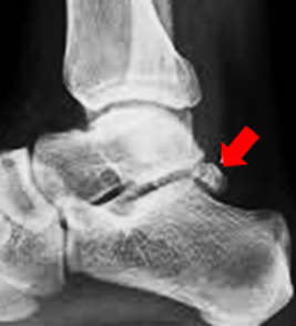
Case study: A 46-Year-Old Woman with Bilateral Foot Pain After a Fall
Acute Jones Fracture
History and Imaging Findings
-
A 46-year-old woman presented with persistent pain in both feet following a fall.
-
Radiographic examination was performed.

Quiz 1:
-
What is the most significant abnormal finding?
(1) Calcaneal stress fracture
(2) Charcot joint
(3) Colles fracture
(4) Jones fracture -
Which of the above is most closely associated with a decrease in Bohler’s angle?
(1) Calcaneal stress fracture
(2) Charcot joint
(3) Colles fracture
(4) Jones fracture -
Where is this fracture located?
(1) Metaphyseal-diaphyseal junction
(2) Diaphysis
(3) Metaphysis
(4) Epiphysis -
What is the final key characteristic of this eponymous fracture?
(1) Extension into the tarso-metatarsal articulation
(2) Extension into the metatarsal-metatarsal articulation
(3) Transverse trajectory
(4) Significant displacement
Follow-up CT Scan
A CT scan of the lower extremity was performed to evaluate for possible narrowing of Bohler’s angle.
|
Böhler’s Angle is measured on a lateral X-ray and represents the angle formed between two lines drawn across specific landmarks of the calcaneus:
The angle formed between these two lines is referred to as Böhler’s angle. 
Böhler’s angle typically falls within the normal range of 20° to 40°. An angle less than 20° is suggestive of a calcaneal fracture. |

No evidence of a calcaneal stress fracture is seen, but the Jones fracture is again identified.
|
A Jones fracture is a fracture that occurs at the base of the fifth metatarsal of the foot, typically involving the bone located on the outer side of the foot.  |
Findings and Diagnosis
Imaging Findings:
Radiographs and CT scans reveal an acute, non-displaced transverse fracture at the base of the fifth metatarsal, extending into the articulation between the fourth and fifth metatarsals.
Narrowing of Böhler’s angle is noted.
An acute dorsal avulsion fracture of the navicular bone is also present.
Soft tissue swelling is observed along the dorsum and lateral aspect of the foot.
Differential Diagnosis:
-
Proximal fifth metatarsal fracture (Zone 1 or Zone 3)
-
Acute Jones fracture (Zone 2)
-
Calcaneal stress fracture
-
Accessory bone (os peroneum)
|
Os Peroneum Definition: Function: Associated Conditions:
 |
Diagnosis:
Acute Jones Fracture (Zone 2)
Discussion
Jones Fracture
Proximal fractures of the fifth metatarsal are classified into three zones: Zone 1, Zone 2, and Zone 3.
A Jones fracture is defined as a fracture of the proximal fifth metatarsal that extends into the articulation between the fourth and fifth metatarsals, corresponding to Zone 2. This type of fracture has a high risk of nonunion.
Pathophysiology
Jones fractures are thought to occur due to a significant adduction force on the forefoot when the ankle is in plantar flexion.
Strong soft tissue attachments to the fifth metatarsal restrict mobility and can lead to displacement or dislocation during injury.
Epidemiology
Fractures of the fifth metatarsal account for approximately 68% of all metatarsal fractures.
Among proximal fifth metatarsal fractures:
-
Zone 1 fractures account for 93%
-
Zone 2 (Jones) fractures account for 4%
-
Zone 3 fractures account for 3%
Clinical Presentation
Patients typically present with lateral foot pain, soft tissue swelling, and difficulty bearing weight, usually following trauma or ankle injury.
Management
Non-displaced fractures of the fifth metatarsal are generally managed non-operatively.
Due to the high risk of nonunion, Jones fractures should be treated with non–non-weight-bearing immobilization using a cast for 6 to 8 weeks.
Reference
(1)
Bowes J, Buckley R. Fifth metatarsal fractures and current treatment. World
J Orthop. 2016;7(12):793-800.
(2)
Chuckpaiwong B, Queen RM, Easley ME, Nunley JA. Distinguishing Jones and
proximal diaphyseal fractures of the fifth metatarsal. Clin Orthop Relat Res.
2008;466(8):1966-1970.
(3)
Metzl JA, Bowers MW, Anderson RB. Fifth metatarsal Jones fractures:
Diagnosis and treatment. J Am Acad Orthop Surg. 2022;30(4):e470-e479.


Comments
Post a Comment